

What Happens During Prophase 1
Prophase is the first step of jail cell partition in mitosis. As it occurs after G2 of interphase, Dna has been already replicated when prophase begins.[1]

Prophase (from Ancient Greek προ- (pro-) 'before', and φάσις (phásis) 'appearance') is the outset stage of prison cell division in both mitosis and meiosis. Kickoff afterwards interphase, DNA has already been replicated when the jail cell enters prophase. The main occurrences in prophase are the condensation of the chromatin reticulum and the disappearance of the nucleolus.[3]
Staining and microscopy [edit]
Microscopy tin can be used to visualize condensed chromosomes as they move through meiosis and mitosis.[4]
Various Deoxyribonucleic acid stains are used to care for cells such that condensing chromosomes can be visualized equally the motion through prophase.[iv]
The giemsa K-banding technique is ordinarily used to identify mammalian chromosomes, merely utilizing the technology on establish cells was originally difficult due to the loftier caste of chromosome compaction in plant cells.[v] [4] One thousand-banding was fully realized for plant chromosomes in 1990.[6] During both meiotic and mitotic prophase, giemsa staining tin can be practical to cells to elicit Grand-banding in chromosomes.[ii] Argent staining, a more modern technology, in conjunction with giesma staining can be used to paradigm the synaptonemal complex throughout the various stages of meiotic prophase.[seven] To perform M-banding, chromosomes must be fixed, and thus information technology is not possible to perform on living cells.[8]
Fluorescent stains such as DAPI can be used in both live plant and animal cells. These stains practise not band chromosomes, but instead allow for DNA probing of specific regions and genes. Use of fluorescent microscopy has vastly improved spatial resolution.[9]
Mitotic prophase [edit]
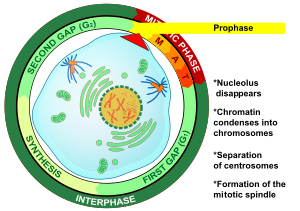
Prophase is the outset stage of mitosis in animal cells, and the second stage of mitosis in plant cells.[ten] At the start of prophase there are two identical copies of each chromosome in the cell due to replication in interphase. These copies are referred to as sis chromatids and are attached by Deoxyribonucleic acid element called the centromere.[xi] The master events of prophase are: the condensation of chromosomes, the movement of the centrosomes, the formation of the mitotic spindle, and the offset of nucleoli break down.[3]
Condensation of chromosomes [edit]
DNA that was replicated in interphase is condensed from DNA strands with lengths reaching 0.7 μm downwardly to 0.2-0.3 μm.[3] This process employs the condensin complex.[11] Condensed chromosomes consist of two sister chromatids joined at the centromere.[12]
Movement of centrosomes [edit]
During prophase in fauna cells, centrosomes movement far plenty apart to exist resolved using a light microscope.[3] Microtubule activity in each centrosome is increased due to recruitment of γ-tubulin. Replicated centrosomes from interphase move apart towards reverse poles of the cell, powered by centrosome associated motor proteins.[thirteen] Interdigitated interpolar microtubules from each centrosome interact with each other, helping to move the centrosomes to contrary poles.[13] [three]
Germination of the mitotic spindle [edit]
Microtubules involved in the interphase scaffolding break down as the replicated centrosomes split.[iii] The movement of centrosomes to opposite poles is accompanied in animal cells past the organisation of individual radial microtubule arrays (asters) by each centromere.[thirteen] Interpolar microtubules from both centrosomes collaborate, joining the sets of microtubules and forming the basic structure of the mitotic spindle.[13] Institute cells do not have centrosomes and the chromosomes can nucleate microtubule assembly into the mitotic apparatus.[xiii] In plant cells, microtubules gather at opposite poles and brainstorm to course the spindle apparatus at locations chosen foci.[x] The mitotic spindle is of swell importance in the process of mitosis and will somewhen segregate the sister chromatids in metaphase.[3]
Get-go of nucleoli breakdown [edit]
The nucleoli brainstorm to break downwards in prophase, resulting in the discontinuation of ribosome production.[3] This indicates a redirection of cellular energy from general cellular metabolism to cellular division.[three] The nuclear envelope stays intact during this process.[ten]
Meiotic prophase [edit]
Meiosis involves ii rounds of chromosome segregation and thus undergoes prophase twice, resulting in prophase I and prophase II.[12] Prophase I is the most complex phase in all of meiosis because homologous chromosomes must pair and exchange genetic data.[3] : 98 Prophase II is very like to mitotic prophase.[12]
Prophase I [edit]
Prophase I is divided into 5 phases: leptotene, zygotene, pachytene, diplotene, and diakinesis. In addition to the events that occur in mitotic prophase, several crucial events occur within these phases such every bit pairing of homologous chromosomes and the reciprocal exchange of genetic material between these homologous chromosomes. Prophase I occurs at different speeds dependent on species and sex. Many species arrest meiosis in diplotene of prophase I until ovulation.[3] : 98 In humans, decades tin can pass as oocytes remain arrested in prophase I only to quickly complete meiosis I prior to ovulation.[12]
Leptotene [edit]
In the first stage of prophase I, leptotene (from the Greek for "delicate"), chromosomes begin to condense. Each chromosome is in a haploid country and consists of ii sister chromatids; however, the chromatin of the sister chromatids is not yet condensed enough to be resolvable in microscopy.[3] : 98 Homologous regions within homologous chromosome pairs begin to associate with each other.[2]
Zygotene [edit]
In the second phase of prophase I, zygotene (from the Greek for "conjugation"), all maternally and paternally derived chromosomes have constitute their homologous partner.[3] : 98 The homologous pairs then undergo synapsis, a procedure by which the synaptonemal complex (a proteinaceous structure) aligns corresponding regions of genetic data on maternally and paternally derived not-sister chromatids of homologous chromosome pairs.[3] : 98 [12] The paired homologous chromosome bound by the synaptonemal complex are referred to every bit bivalents or tetrads.[x] [iii] : 98 Sex (X and Y) chromosomes do not fully synapse because only a modest region of the chromosomes are homologous.[three] : 98
The nucleolus moves from a central to a peripheral position in the nucleus.[14]
Pachytene [edit]
The third phase of prophase I, pachytene (from the Greek for "thick"), begins at the completion of synapsis.[3] : 98 Chromatin has condensed enough that chromosomes can now be resolved in microscopy.[ten] Structures chosen recombination nodules course on the synaptonemal circuitous of bivalents. These recombination nodules facilitate genetic substitution between the not-sis chromatids of the synaptonemal complex in an effect known as crossing-over or genetic recombination.[3] : 98 Multiple recombination events tin occur on each bivalent. In humans, an average of 2-three events occur on each chromosome.[13] : 681
Diplotene [edit]
In the fourth phase of prophase I, diplotene (from the Greek for "twofold"), crossing-over is completed.[3] : 99 [10] Homologous chromosomes retain a full prepare of genetic information; all the same, the homologous chromosomes are now of mixed maternal and paternal descent.[3] : 99 Visible junctions called chiasmata agree the homologous chromosomes together at locations where recombination occurred equally the synaptonemal complex dissolves.[12] [3] : 99 It is at this stage where meiotic arrest occurs in many species.[3] : 99
Diakinesis [edit]
In the fifth and final phase of prophase I, diakinesis (from the Greek for "double movement"), full chromatin condensation has occurred and all iv sister chromatids tin can be seen in bivalents with microscopy. The balance of the phase resemble the early stages of mitotic prometaphase, as the meiotic prophase ends with the spindle apparatus beginning to grade, and the nuclear membrane starting time to break downwards.[x] [3] : 99
Prophase II [edit]
Prophase Two of meiosis is very similar to prophase of mitosis. The most noticeable difference is that prophase 2 occurs with a haploid number of chromosomes as opposed to the diploid number in mitotic prophase.[12] [10] In both animal and plant cells chromosomes may de-condense during telophase I requiring them to re-condense in prophase II.[3] : 100 [ten] If chromosomes exercise not need to re-condense, prophase II often proceeds very quickly as is seen in the model organism Arabidopsis.[10]
Prophase I arrest [edit]
Female person mammals and birds are born possessing all the oocytes needed for future ovulations, and these oocytes are arrested at the prophase I phase of meiosis.[xv] In humans, as an example, oocytes are formed between three and four months of gestation within the fetus and are therefor present at birth. During this prophase I arrested stage (dictyate), which may terminal for decades, four copies of the genome are nowadays in the oocytes. The adaptive significance of prophase I arrest is even so not fully understood. Withal, it has been proposed that the arrest of ooctyes at the four genome re-create stage may provide the informational redundancy needed to repair damage in the DNA of the germline.[15] The repair process used appears to exist homologous recombinational repair[fifteen] [16] Prophase arrested oocytes have a loftier capability for efficient repair of Deoxyribonucleic acid amercement.[16] DNA repair capability appears to exist a key quality control mechanism in the female germ line and a critical determinant of fertility.[16]
Differences in plant and creature cell prophase [edit]

Arabidopsis thaliana prison cell in preprophase, prophase and prometaphase. Preprophase band is present along the cell wall from images ane–3, is fading in image four, and disappears by paradigm five.[1]
The most notable difference betwixt prophase in constitute cells and animal cells occurs because plant cells lack centrioles. The organization of the spindle apparatus is associated instead with foci at opposite poles of the cell or is mediated by chromosomes. Another notable difference is preprophase, an additional step in constitute mitosis that results in formation of the preprophase band, a structure equanimous of microtubules. In mitotic prophase I of plants, this band disappears.[x]
Prison cell checkpoints [edit]
Prophase I in meiosis is the virtually complex iteration of prophase that occurs in both plant cells and animal cells.[3] To ensure pairing of homologous chromosomes and recombination of genetic fabric occurs properly, in that location are cellular checkpoints in place. The meiotic checkpoint network is a Deoxyribonucleic acid impairment response system that controls double strand interruption repair, chromatin structure, and the movement and pairing of chromosomes.[17] The organisation consists of multiple pathways (including the meiotic recombination checkpoint) that prevent the cell from entering metaphase I with errors due to recombination.[xviii]
Encounter also [edit]
- Metaphase
- Anaphase
- Telophase
- Meiosis
- Mitosis
- Cytoskeleton
- Homologous chromosome
References [edit]
- ^ a b Nussbaum RL, McInnes RR, Huntington F (2016). Thompson & Thompson Genetics in Medicine. Philadelphia: Elsevier. pp. 12–20. ISBN9781437706963.
- ^ a b c Schermelleh L, Carlton PM, Haase S, Shao L, Winoto L, Kner P, et al. (June 2008). "Subdiffraction multicolor imaging of the nuclear periphery with 3D structured illumination microscopy". Science. 320 (5881): 1332–36. Bibcode:2008Sci...320.1332S. doi:10.1126/scientific discipline.1156947. PMC2916659. PMID 18535242.
- ^ a b c d eastward f g h i j thou l m northward o p q r s t u v w x y Hartwell LH, Hood Fifty, Goldberg ML, Reynolds AE, Silver LM, Veres RC (2008). Genetics From Genes to Genomes . New York: McGraw-Hill. pp. 90–103. ISBN978-0-07-284846-5.
- ^ a b c Singh RJ (2017). Plant Cytogenetics (3rd ed.). Boca Raton, FL: CBC Press, Taylor & Francis Group. p. xix. ISBN9781439884188.
- ^ Wang HC, Kao KN (1988). "G-banding in plant chromosomes". Genome. 30: 48–51. doi:10.1139/g88-009 – via ResearchGate.
- ^ Kakeda G, Yamagata H, Fukui K, Ohno Thou, Fukui Grand, Wei ZZ, Zhu ES (Baronial 1990). "High resolution bands in maize chromosomes by G-banding methods". Theoretical and Applied Genetics. 80 (2): 265–72. doi:10.1007/BF00224397. PMID 24220906. S2CID 6600449.
- ^ Pathak S, Hsu TC (Jan 1979). "Argent-stained structures in mammalian meiotic prophase". Chromosoma. 70 (2): 195–203. doi:x.1007/bf00288406. PMID 85512. S2CID 27763957.
- ^ Sumner AT (May 1982). "The nature and mechanisms of chromosome banding". Cancer Genetics and Cytogenetics. 6 (one): 59–87. doi:10.1016/0165-4608(82)90022-x. PMID 7049353.
- ^ de Jong H (December 2003). "Visualizing DNA domains and sequences by microscopy: a fifty-yr history of molecular cytogenetics". Genome. 46 (six): 943–half-dozen. doi:x.1139/g03-107. PMID 14663510.
- ^ a b c d e f g h i j k Taiz L, Zeiger East, Moller IM, Spud A (2015). Plant Physiology and Development. Sunderland MA: Sinauer Associates. pp. 35–39. ISBN978-1-60535-255-8.
- ^ a b Zeng XL, Jiao MD, Wang XG, Vocal ZX, Rao Southward (2001). "Electron microscopic studies on the Silvery-stained Nucleolar Cycle of Physarum Polycephalum" (PDF). Acta Botanica Sinica. 43 (seven): 680–5. Archived from the original (PDF) on 2018-x-01. Retrieved 24 February 2015.
- ^ a b c d east f g Nussbaum RL, McInnes RR, Willard HF (2016). Thompson & Thompson Genetics in Medicine. Philadelphia: Elsevier. pp. 12–20. ISBN978-one-4377-0696-three.
- ^ a b c d e f Alberts B, Bray D, Hopkin 1000, Johnson A, Lewis J, Raff 1000, Roberts K, Walter P (2004). Essential Cell Biological science. New York NY: Garland Science. pp. 639–658. ISBN978-0-8153-3481-i.
- ^ Zickler D, Kleckner N (1998). "The leptotene-zygotene transition of meiosis". Annual Review of Genetics. 32: 619–97. doi:10.1146/annurev.genet.32.1.619. PMID 9928494.
- ^ a b c Mira A (September 1998). "Why is meiosis arrested?". Journal of Theoretical Biology. 194 (2): 275–87. Bibcode:1998JThBi.194..275M. doi:10.1006/jtbi.1998.0761. PMID 9778439.
- ^ a b c Stringer JM, Winship A, Zerafa N, Wakefield M, Hutt Yard (May 2020). "Oocytes can efficiently repair Dna double-strand breaks to restore genetic integrity and protect offspring health". Proceedings of the National Academy of Sciences of the United states. 117 (21): 11513–11522. doi:ten.1073/pnas.2001124117. PMC7260990. PMID 32381741.
- ^ Hochwagen A, Amon A (March 2006). "Checking your breaks: surveillance mechanisms of meiotic recombination". Current Biology. 16 (half-dozen): R217-28. doi:10.1016/j.cub.2006.03.009. PMID 16546077.
- ^ MacQueen AJ, Hochwagen A (July 2011). "Checkpoint mechanisms: the boob masters of meiotic prophase". Trends in Jail cell Biological science. 21 (7): 393–400. doi:10.1016/j.tcb.2011.03.004. PMID 21531561.
External links [edit]
-
 Media related to Prophase at Wikimedia Commons

What Happens During Prophase 1,
Source: https://en.wikipedia.org/wiki/Prophase
Posted by: ablerithey.blogspot.com
0 Response to "What Happens During Prophase 1"
Post a Comment